Kienböck’s disease is a problem in the wrist caused by the loss
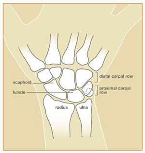
of blood supply to the lunate. The lunate is one of the eight small
bones that make up the “carpal bones” in the wrist (see
Figure 1). There are two rows of bones: one closer to the forearm, the
“proximal row;” the other closer to the fingers, the
“distal row.” The lunate bone is in the center of the
proximal row. It is next to the scaphoid bone, which spans the two
rows.
There is probably no single cause for loss of blood supply to the
lunate. The cause of Kienböck’s disease seems to involve multiple
factors. These factors include the blood supply (arteries), the blood
drainage (veins), and skeletal variations. Skeletal variations
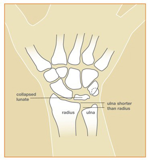
associated with Kienböck’s disease include a shorter length of the
ulna, one of the forearm bones, and the shape of the lunate bone itself
(see Figure 2). There may be some cases that are associated with
diseases like gout, sickle cell anemia, and cerebral palsy.
Trauma, either a single event or repeated significant trauma, may
affect the blood supply to the lunate. In general, though,
Kienböck’s disease is not felt to be related to occupational
hazards. However, the presence of Kienböck’s disease can affect
the treatment and prognosis for traumatic events.
Most patients with Kienböck’s disease present with wrist pain.
There is usually tenderness directly over the lunate bone. The diagnosis
of Kienböck’s disease is made by history, physical examination,
and plain x-rays. Special studies are sometimes also needed to confirm
the diagnosis. Probably the most reliable special study to assess the
blood supply of the lunate is Magnetic Resonance Imaging, or MRI. CT
scanning, specialized CT scanning, and bone scan may also be
used.
The progression of Kienböck’s disease is variable and
unpredictable. Sometimes the disease may be diagnosed at a very early
stage. At this time, there may be only pain and swelling, and normal
x-rays. As the disease progresses the x-ray changes in the lunate become
more obvious. With further progression, the lunate develops small
fractures and the bone fragments and collapses. As collapse occurs, the
mechanics of the wrist become changed, which puts abnormal stresses and
wear on the joints within the wrist itself. One should be aware that not
every case of Kienböck’s disease progresses through all stages to
the severely deteriorated arthritic end-stage.
Treatment options depend upon the severity and stage of the disease.
In very early stages, the treatment can be as simple as observation or
immobilization. For more advanced stages, surgery is usually considered
to try to reduce the load on the lunate bone by lengthening, shortening,
or fusing various bones in the forearm or wrist.Sometimes bone grafting
or removal of the diseased lunate is performed. If the disease is very
advanced and the relations of the bones one to the other have markedly
deteriorated, complete wrist fusion may be the treatment of
choice.
Hand therapy does not change the course of the disease; however, hand
therapy can help to minimize the disability from the problem. Treatment
is designed to relieve pain and restore function.
Your hand surgeon will advise you of the best treatment.Your hand
surgeon can explain the risks, benefits, and side-effects of various
treatments for Kienböck’s disease.
The results of Kienböck’s disease and its treatment vary
considerably depending on the severity of the involvement, and whether
or not the disease progresses. The disease process and response to
treatment will take several months. On occasion, several forms of
treatment, and even multiple operations, might be necessary.

Figure 1: Normal wrist

Figure 2: Wrist with Kienböck’s disease and ulna that is
short compared to radius
© 2006 American Society for Surgery of the Hand


