The hand is known to frequently present signs or symptoms of generalized or systemic diseases. Because the hand contains structures from the skin, muscular, skeletal, circulatory, and nervous systems, it often provides clues to diseases which are yet to be diagnosed in other parts of the body. Presentation of complaints, lesions, symptoms or signs in the hand may alert the hand surgeon to these systemic diseases. Although the hand surgeon may not treat all of these diseases, he is in a position to help make the diagnosis and refer the patient to a different specialist.
Musculoskeletal diseases may result in enlargement of the joints. Arthritis diseases commonly are associated with these abnormalities.
Enlargement of the middle joint of a finger. This is called a Bouchard’s Node.
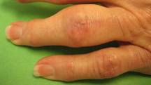
Figure 1: This is a common finding in osteoarthritis. There is often pain and stiffness. Deformity is also seen.
Enlargement of the small joint of a finger or thumb is called a Heberden’s node.
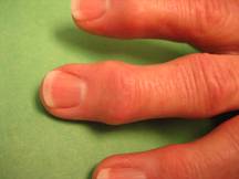
Figure 2: This is a classic sign in osteoarthritis. There may or not be pain, but stiffness or instability may be present. Deformity to the side or down is common when more severe.
Enlargement of a finger/thumb joint may also occur from deposition of crystals from diseases like Gout.
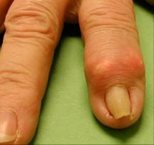
Figure 3: The overlying skin and even skin away from the joint may be discolored whitish or yellow from the deposits of urate salt in the tissues. If the overlying skin becomes too thin, the whitish-yellow salts may drain through the skin, simulating a ruptured boil or abscess. The ruptured areas can become secondarily infected.
Enlargement of a whole digit, associated with inflammation, may be due to an inflammatory disease. One form is called dactylitis.
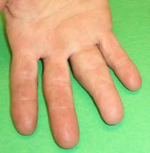
Figure 4: This case of dactylitis was associated with psoriatic arthritis. In this photo, the swelling extends from the palm to include the ring finger out to the small joint. This is commonly associated with stiffness. Pain may also be present. The condition may respond to medical treatment for the underlying condition.
Small localized cystic swellings, especially at the small finger/thumb joints, can be associated with osteoarthritis.
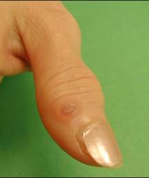
Figure 5: This type of cyst is called a mucous cyst or ganglion cyst. There may or may not be pain at the underlying arthritic joint, although the arthritis is considered the cause. If the skin becomes thin, spontaneous rupture may occur, resulting in drainage of a clear sticky fluid. Although not in itself a problem, this rupture may allow bacteria to reach the adjacent joint, causing joint or bone infection.
Small hemorrhages in the cuticle or dilation of the small vessels have been seen in musculoskeletal diseases.
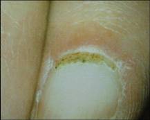
Figure 6: The small red dots may be seen, as in this photo, at the thinnest portion, but can also be seen in the pink portion of the cuticle. This has been seen in dermatomyositis, systemic lupus, and scleroderma.
Many different diseases may affect the fingernails. Changes may occur in coloration, shape, curvature, smoothness, or attachment to the underlying nail bed. White coloration may affect the nail bed itself or may be caused by changes in the nail. Leukonychia is a term used to describe white color of the nail.
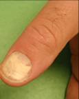
Figure 7: Leukonychia has been reported with viral infections, intestinal and kidney diseases, poisoning, medications, and detachment of the nail from the nail bed by fungal infections & other causes.
Apparent leukonychia is a term used to describe white coloration due to changes deep to the nail itself or detachment of the nail (onycholysis).
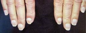
Figure 8: Half & Half nails have been associated with chronic kidney failure.
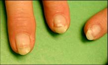
Figure 9: Terry’s nails are white from the cuticle out to nearly the end of the nail bed, which retains a normal pink color, and are seen in liver and heart.
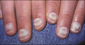
Figure 10: Muehrche’s nails have lines of white color transversely through the nail bed. Low serum albumin (blood protein) and kidney failure are two causes.
Red, blue, grey, black, and yellow colorations have also been reported.
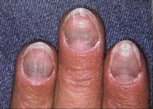
Figure 11: The blue color beyond the base of the fingernails in this patient was associated with the use of a medication called minocycline.
Yellow colored nails have been associated with medications, and diseases of the lung, liver, and sinuses.
Diseases of the heart and blood vessels have been associated with a number of different manifestations in the nails.
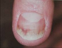
Figure 12: Red streaks seen in the fingernail area can be due to hemorrhage (bleeding). These are called splinter hemorrhages and have been seen in endocarditis (heart infection), although also reported in psoriasis, and trichinosis.
Clubbed nails have an abnormal curvature from the cuticle to tip and from side to side.
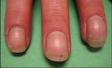
Figure 13: Clubbed nails have been described in three situations. Although these can be hereditary or congenital, they have greater importance if acquired, as this could be a sign of heart and/or lung disease.
Breakdown of the tissues at the tips of the fingers can be a sign of underlying disease of the arteries.
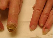
Figure 14: This patient suffers from Buerger’s disease, which causes slow but progressive deterioration of circulation. The tissues become gangrenous and amputation is often necessary as the disease is difficult to treat/reverse.
Other diseases of blood vessels may be present for years and cause little difficulty, such as temporary bluish or purple color due to poor circulation.
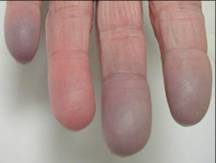
Figure 15: Raynaud’s disease or phenomenon (temporary spasm of the arteries in the fingers) has been reported by itself, but also with rheumatoid arthritis, systemic lupus, and scleroderma, to name a few. If the condition is more severe, fingertip ulcers from the poor circulation may occur. Sometimes medication may be helpful.
As noted above, some medications have effects upon the tissues of the fingers. In addition to color changes (blue, yellow, brown, etc.) of the nail, the skin can be affected.
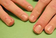
Figure 16: Redness, swelling, and tenderness of the cuticles near the fingertip, followed by the appearance of moist, oozing and easily bleeding tissue (pyogenic granuloma) is a reported side effect of an acne medication, isotretinoin.
Skin diseases are a known cause of some nail abnormalities.
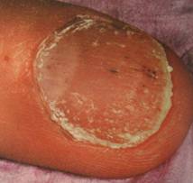
Figure 17: Psoriasis commonly affects the nail and matrix. Pitting (seen here), detachment from the nail bed, nail bed hemorrhages, and other changes may occur.
Other changes in the shape of the fingernails may signal systemic disease.

Figure 18: This “Pincer” nail with an exaggerated side to side curvature can be congenital or simply due to advancing age. However, it can be due, when acquired, to an enlargement of the underlying bone from disease (gout in this case).
Systemic neurological conditions are known to cause many symptoms in the hand and fingers. Systemic conditions most often cause diffuse abnormal sensations (frequently hands and feet) as opposed to entrapment of nerves in the neck/elbow/wrist/hand, which are associated with abnormal sensation localized to the areas specifically served by those individual nerves. Some of the diseases which have been associated with diffuse upper limb numbness/tingling are:
1. |
Diabetic neuropathy
|
2. |
Alcoholic neuropathy
|
3. |
Neuropathy associated with blood vessel spasm diseases:
Rheumatoid arthritis
Systemic Lupus Erythematosis
Polyarteritis Nodosa
|
4. |
Multiple Sclerosis
|
Numbness/tingling in the upper limb (sometimes upper and lower) have been associated with other diseases and with medications. A few are listed below:
1. |
Myofascial Pain Syndrome (tender points in muscles which are often at a distance from the areas of numbness/tingling). The location of symptoms may vary on a daily basis and may include fatigue, “weakness”, clumsiness, heaviness.
|
2. |
Vitamin B12 deficiency syndrome
|
3. |
Therapeutic drugs:
Taxol, Vincristine, Gold,
Cis-platinum, Isoniazid, Pyridoxine
|
4. |
Others:
Crohn’s disease, Amyloidosis, Multiple Myeloma
Dysproteinemia, Arsenic poisoning
|
Triggering action of fingers during flexion and extension (trigger fingers) has also been linked to diabetes mellitus.
Thomas R. Boyce, MD
Clinical Assoc. Professor
Dept. of Orthopaedics and Sports Medicine
University of Washington School of Medicine
© 2006 American Society for Surgery of the Hand